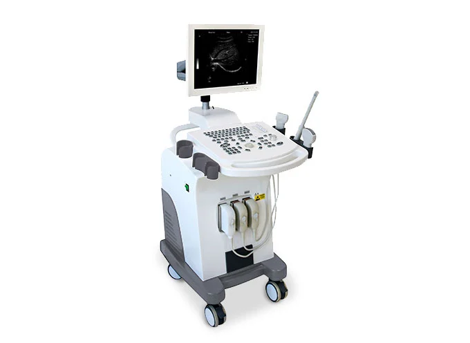
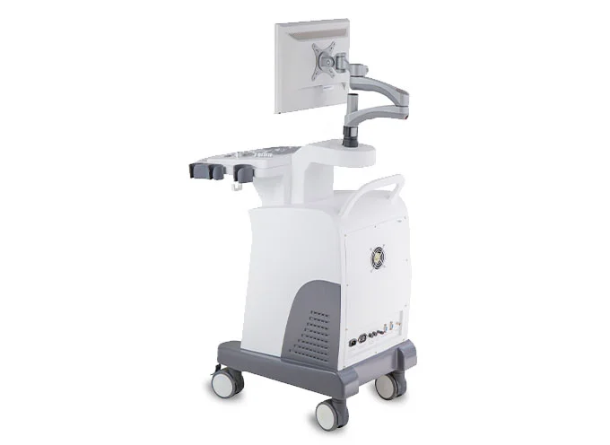
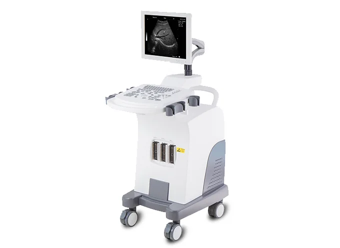
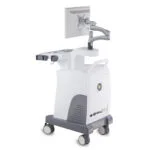
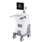
Sonoview® Fully Digital Ultrasound Imaging System with clear image, PC-based, extensive functions, 15-inch color LCD screen, multi-frequency broadband probes, upgradeable to 3D
All Meditech customers and healthcare professionals can submit equipment service tickets through the Customer Service Portal.
To receive two additional months of extended warranty, please register your product here.To browse all support resources and contact information, please visit the Support Center.
Coming soon.
Discover the advanced capabilities that set our products apart
Standard configuration:
Options:
Detailed technical specifications and performance parameters
| Category | Specifications |
|---|---|
| Display | 14-inch monochrome SVGA display with Progressive Scan technology |
| Scanning Depth | Up to 240 mm |
| Lateral Resolution | 2 mm |
| Axial Resolution | 1 mm |
| Grayscale | 256 levels |
| TGC Settings | 8 settings |
| Imaging Modes | B, 2B, B/M, M, 4B, Zoom |
| Zoom | Real-time zoom in B mode, at ratios of 1.0×, 1.5×, 2.0×, 2.5×, 3.0×, 3.5×, 4.0×, 4.5×, 5.0×, 5.5×, 6.0× |
| 3D Imaging | Optional 3D |
| Main Probe | Electronic convex probe 3.5 MHz |
| Frequency Switching | 5 frequencies |
| Electronic Focus | 4 electronic foci with the ability to set focus positions |
| Image Processing | Pre-processing and post-processing, dynamic range, frame rate, midline and edge enhancement, image inversion, grayscale adjustment, contrast, brightness and Gamma correction |
| Image Orientation | Up, down, right and left |
| Pseudo Colors | 8 patterns |
| Cine Loop | 64, 128, 256, 512, 1024 frames with the ability to preset the capacity |
| Body Marks | Approximately 30 types of body marks |
| Display Information | Date, time, medical record number, zoom, measurement values, body marks, comments, image correlation coefficient, scanning depth and probe type |
| Text Editing | Full-screen text editing |
| Languages | Supports English and Chinese |
| B Mode Measurements | Distance, circumference, area, volume, angle, ratio, stenosis, curve and histogram |
| M Mode Measurements | Heart rate, time, distance, slope and stenosis |
| Gynecology Measurements | Uterus, cervix, endometrium, right and left ovary |
| Obstetrics Measurements | Gestational age, fetal weight, amniotic fluid index AFI |
| Cardiac Measurements | LV, left ventricular function, LVPW, RVAWT |
| Urology Measurements | Transitional zone, bladder volume, RUV, prostate and kidneys |
| Small Parts Measurements | Eye, thyroid, maxillofacial |
| DICOM | Supports DICOM 3.0 for transferring medical images and data and integration with PACS systems |
| Presets | Gynecology, obstetrics, vascular, cardiac, urology and small parts |
| Storage Memory | High-capacity local memory with USB support |
| Saved Data | Images, Cine Loop, measurement results and reports |
| Probe Ports | 2 ports for connecting probes |
| Video Outputs | VGA and PAL-D |
| Printing | Support for laser printers and inkjet printers |
| DVD-ROM | Optional DVD-ROM reader/writer |
| Dimensions | 790 × 660 × 1070 mm |
| Weight | Approximately 50 kg |
Compatible accessories to extend functionality
| Item | Quantity |
|---|---|
| Mobile Trolley | 1 Piece |
| Multi-frequency Convex Probe | 1 Piece |
| Power Cable | 1 Piece |
| Cleaning Cloth | 1 Piece |
| Item |
|---|
| High-frequency Linear Array Probe 7.5 MHz |
| Intracavity (Transvaginal) Probe 6.5 MHz |
| Transrectal Probe 7.5 MHz |
| Small Convex Probe 3.5 MHz |
| Reader/Writer DVD-ROM |
| Laser Printer Black and White (B/W) |
Core specifications at a glance
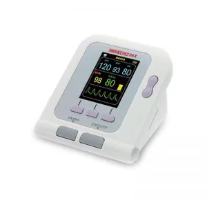
Portable patient monitor NIBP and SpO₂, a reliable and integrated vital signs monitoring device designed for accurate measurement of non-invasive blood pressure, oxygen saturation (SpO₂), and pulse rate.
Learn more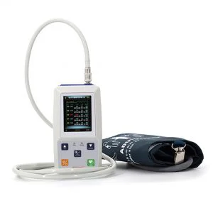
The ambulatory blood pressure monitoring device combines continuous blood pressure monitoring and oxygen saturation in a compact design.
Learn more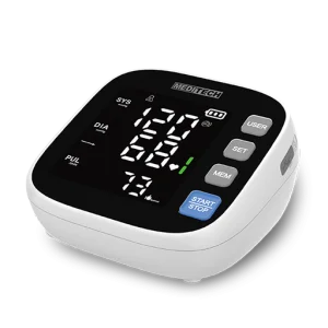
The smart digital blood pressure monitor provides accurate and convenient blood pressure monitoring with a large LED display, bilingual voice guidance, and memory storage for three users.
Learn more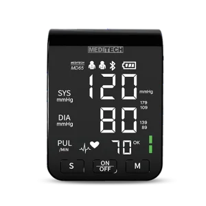
The digital blood pressure monitor is an advanced and easy-to-use device designed for accurate measurement of blood pressure...
Learn more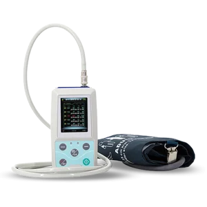
The ambulatory blood pressure monitoring device for outpatients is a compact and easy-to-use device designed for continuous 24-hour blood pressure monitoring.
Learn more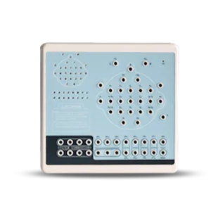
The 32-channel EEG Brain Mapping System is an advanced digital electroencephalography device designed for comprehensive brain activity monitoring and neurological diagnosis.
Learn more